Cross Section Of A Bone : Exam 1 - Animal Sciences 511 with Berndtson at University .... This is known as the periosteum. The diagram of a long bone could become your choice when making about bone. I don't find it enhances the image. Browse 4,253 bone cross section stock photos and images available, or search for human bone cross section to find more great stock photos and pictures. Related posts of cross section of human bone diagram human back muscles and bones.
(area/long bone length 3) ∗ 10 8. On examining a cross section of any bone, it is composed of two kinds of bony tissue: The cell line involved in osteogenesis consists of preosteoblasts, osteoblasts, osteocytes and bone. The large dark spots are passages for blood vessels and nerves. Browse 4,253 bone cross section stock photos and images available, or search for human bone cross section to find more great stock photos and pictures.
Long Bone Parts and Blood Supply | Bone and Spine from i2.wp.com Decalcified compact bone looks completely different than compact bone that still has calcium salts in its matrix. Sections of bone marrow tissue. Two types of bone tissues in cross section of a long bone : Foot bone anatomy x ray 12 photos of the foot bone anatomy x ray foot bone anatomy x ray, bone, foot bone anatomy x ray. The cell line involved in osteogenesis consists of preosteoblasts, osteoblasts, osteocytes and bone. It consists of two layers; Body size standardization was done, using the following equations: The section has been ground and dried, hence the lacunae… rienstra clinic medicine for people!
The humerus is the long bone located in the upper arm of the body which extends from the shoulder joint to the elbow.
The large dark spots are passages for blood vessels and nerves. In three dimensions an osteon is cylindrical in shape. Bone and bones / pathology*. Concentric layers of bone cells (osteocytes) and bone matrix surround the central canal. Cross section of a femur bone showing the anatomical structure including cancellous bone and marrow. In the center of each osteon is the central canal, a space that houses blood vessels and nerves that supply bone. Each bone in your body is made up of three main types of bone material: Bone cross section for radius digital science on behance. The section has been ground and dried, hence the lacunae… rienstra clinic medicine for people! The humerus is the long bone located in the upper arm of the body which extends from the shoulder joint to the elbow. Browse 53 bone marrow cross section stock photos and images available, or search for bone cross section or bone cells to find more great stock photos and pictures. Histology slide courtesy of william l. Now that you know what bones do, let's take a look at what they're made of and their anatomy.
Now that you know what bones do, let's take a look at what they're made of and their anatomy. Cross section of a bone : Table 1 describes the bone markings, which are illustrated in (figure 4). The surface features of bones vary considerably, depending on the function and location in the body. An outer 'fibrous layer' containing mainly fibroblasts, and an inner 'cambium layer' containing progenitor cells.
Dinosaur Bone Cross Section | Osteon remains of this bone ... from c1.staticflickr.com Two types of bone tissues in cross section of a long bone : And why does the marrow stop where it does, and so sharply? The large dark spots are passages for blood vessels and nerves. Table 1 describes the bone markings, which are illustrated in (figure 4). Related posts of cross section of a long bone foot bone anatomy x ray. To the left is muscle tissue, and to the right Bone matrix and cells bone matrix osseous tissue is a connective tissue and like all connective tissues contains relatively few cells and large amounts of extracellular matrix. The surface features of bones vary considerably, depending on the function and location in the body.
This is known as the periosteum.
The compact bone is made up of osteon. The upper (biting) surfaces of the tooth are at top, with the lower sections (bottom) embedded in the gums and jaw bone (not shown). Smartdraw includes 1000s of professional healthcare and anatomy chart templates that you can modify and make your own. Cross section of the shaft (diaphysis). Would it be a good thing to show the epiphyseal plate? Start studying cross section of bone. Learn vocabulary, terms, and more with flashcards, games, and other study tools. Cross section of mandible at first molar region showing cortical and spongy bone basic concepts in osteogenesis. On examining a cross section of any bone, it is composed of two kinds of bony tissue: Browse 4,253 bone cross section stock photos and images available, or search for human bone cross section to find more great stock photos and pictures. Bone cross section for radius digital science on behance. After a fracture, woven bone forms initially and is gradually replaced by lamellar bone during a process known as bony substitution. Decalcified compact bone looks completely different than compact bone that still has calcium salts in its matrix.
Each bone in your body is made up of three main types of bone material: Human back muscles and bones 12 photos of the human back muscles and bones human back muscles and bones, bone, human back muscles and bones This is essentially a measure of how the material is distributed about a given axis. While it is not as hard as compact bone, spongy bone plays an important role of protecting the marrow where blood cells are produced. To the left is muscle tissue, and to the right
Plate 6.21 from www.anatomyatlases.org In addition, cortical bone thickness at anterior, posterior, medial, and lateral parts of the bone section was measured. Bone on side of the foot Two types of bone tissues in cross section of a long bone : Start studying cross section of long bone. Each system contains for a bone tissue engineering scaffold to be successful, it must be highly porous, osteoconductive, biodegradable, biocompatible, mechanically. The surface features of bones vary considerably, depending on the function and location in the body. Foot bone anatomy x ray 12 photos of the foot bone anatomy x ray foot bone anatomy x ray, bone, foot bone anatomy x ray. Bone matrix and cells bone matrix osseous tissue is a connective tissue and like all connective tissues contains relatively few cells and large amounts of extracellular matrix.
I = ∫ (y2 δa).
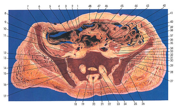
They are obtained by taking imaginary slices perpendicular to the main axis of organs, vessels, nerves, bones, soft tissue, or even the entire human body. Start studying cross section of long bone. Related posts of cross section of a long bone foot bone anatomy x ray. The arrows point toward the tumor. Table 1 describes the bone markings, which are illustrated in (figure 4). Cross section of mandible at first molar region showing cortical and spongy bone basic concepts in osteogenesis. After a fracture, woven bone forms initially and is gradually replaced by lamellar bone during a process known as bony substitution. This slide contained a cross section of a very small bone, and you are looking at the entire thickness of the shaft of the bone. Compact bone cross section courtesy: Each bone in your body is made up of three main types of bone material: Cross section of a bone : I = ∫ (y2 δa). Bone on side of the foot